A New Way to Measure Coronal Spinal Alignment — and Why It Predicts Outcomes Better Than the Standard Approach
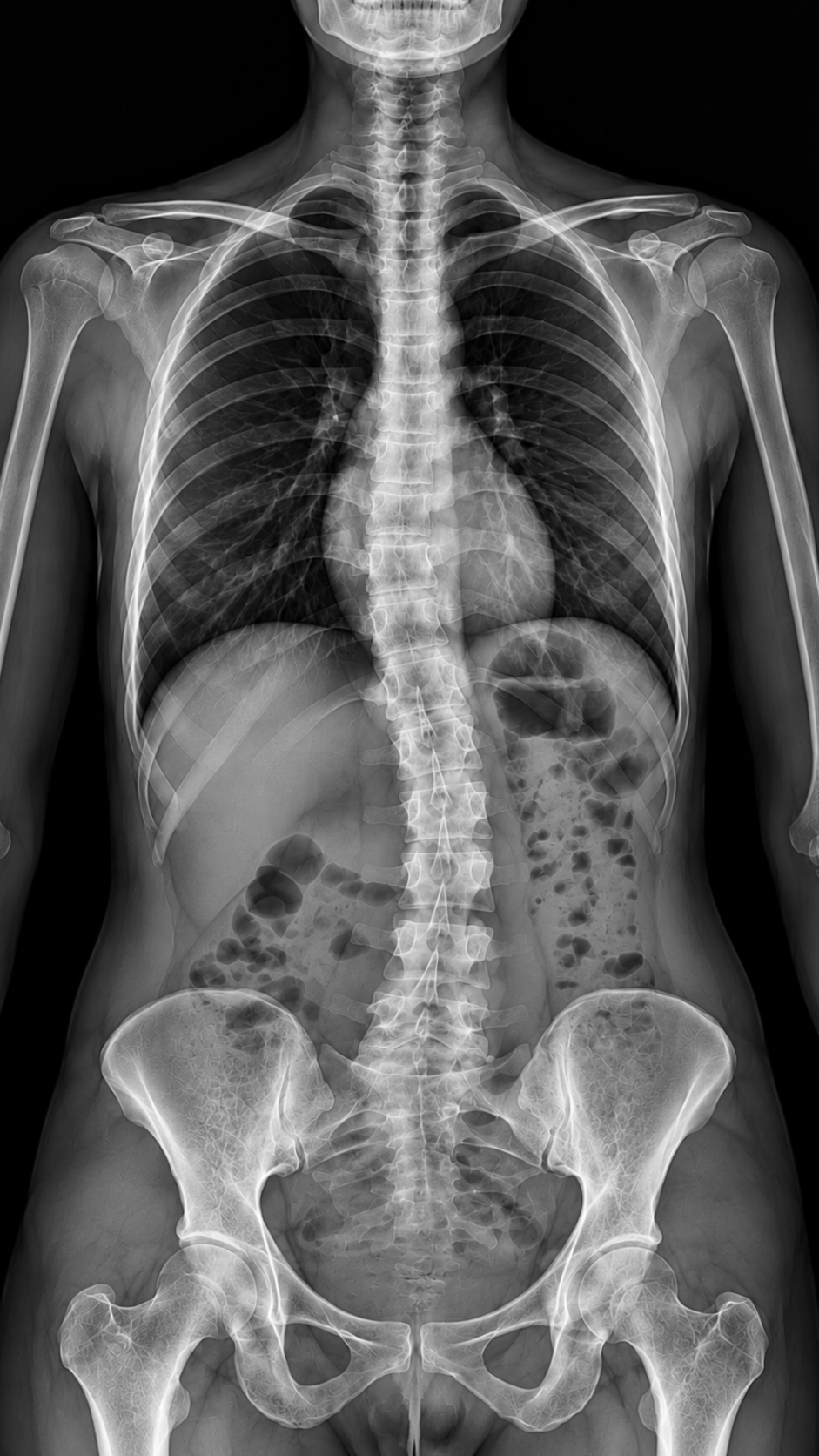
An ISSG multicenter study of 328 patients introduces the C2 coronal angle as a novel angular measure of coronal balance in adult spinal deformity surgery — finding it independently predicts neck disability and quality of life, with a clinically actionable threshold of 3.5°.
OPENING SUMMARY
How we measure something shapes what we learn from it. In adult spinal deformity surgery, coronal alignment — the side-to-side balance of the spine — has traditionally been assessed using a single linear measurement: the horizontal distance between C7 and the central sacral vertical line (C7-CSVL), expressed in millimeters. This approach has been clinically useful, but it has a fundamental limitation: it describes the magnitude of offset at one level of the spine without capturing the angular relationship between the top of the construct and the coronal midline. In the sagittal plane, vertebropelvic angles have long complemented linear measurements to provide a more complete picture of alignment and its clinical consequences. In the coronal plane, no analogous angular measure has existed — until now. This study introduces the C2 coronal angle, defines its optimal clinical threshold, and demonstrates that it predicts patient-reported outcomes more strongly than the linear measure it is designed to complement.
STUDY SNAPSHOT
Study type – Retrospective multicenter analysis
Number of patients – 328
Mean age – 64.0 years
Mean levels fused – 12.6
Surgical procedure – Thoracolumbar instrumentation to the pelvis
Novel metric – C2 coronal angle (C2C) — angle between CSVL and center of C2 body
Comparator – C7-CSVL linear offset
Primary outcome timepoint – 2-year follow-up
Primary outcomes – SRS-22, SF-36, NDI; MCID achievement
Optimal C2C threshold – 3.5° (by ROC / Youden's Index)
OR for neck disability above threshold – 1.90 (P=0.049)
OR reduction in NDI MCID per 1° C2C increase – 34% (OR=0.66; P=0.022)
C2C vs. C7-CSVL model fit for SRS-Total – AIC 657 vs. 660; adj. R² 0.058 vs. 0.048
Published in – Spine, online ahead of print, September 11, 2026
PMID – 42766480
DOI – 10.1097/BRS.0000000000005871
Lead author – Nikita Lakomkin
WHY THIS MATTERS
Coronal alignment — how the spine balances from side to side when viewed from the front — is one of the fundamental dimensions of spinal deformity assessment. Residual coronal imbalance after surgery is associated with pain, disability, and reduced quality of life. Yet the tools used to measure and target coronal alignment have not kept pace with the sophistication of sagittal alignment assessment.
In sagittal planning, the field has embraced angular measures — pelvic tilt, pelvic incidence, T1 pelvic angle, sagittal vertical axis — that capture the geometric relationships between spinal segments and the pelvis in ways that linear offsets cannot. These angular measures have proven more predictive of outcomes than simple linear measurements of sagittal balance. The question this study asks is whether the same logic applies in the coronal plane: would an angular measure of coronal alignment — one that captures the tilt of the entire spinal column relative to the midline rather than just the offset at C7 — better predict what happens to patients after surgery?
The answer, based on this multicenter analysis of 328 patients, is yes. The C2 coronal angle was independently associated with patient-reported outcomes across multiple validated instruments, outperformed the traditional C7-CSVL linear offset in predictive modeling, and yielded a specific threshold — 3.5° — that is immediately applicable as a surgical target.
UNDERSTANDING THE MEASUREMENTS
C7-CSVL (Traditional Linear Measure)
The traditional measure of coronal alignment in ASD surgery is the C7-CSVL offset — the horizontal distance in millimeters between the center of the C7 vertebral body and the central sacral vertical line (CSVL), a plumb line drawn upward from the midpoint of the sacrum. A large offset indicates that the upper spine is displaced to one side of the pelvis, reflecting coronal imbalance. This measurement is straightforward to obtain from standard radiographs and has been widely used in clinical practice and research. Its limitation is that it captures displacement only at the C7 level and does not reflect the angular orientation of the cervical spine relative to the coronal midline.
C2 Coronal Angle (Novel Angular Measure)
The C2 coronal angle measures the angle between the central sacral vertical line and a line connecting the CSVL reference point to the center of the C2 vertebral body. Rather than asking "how far is C7 from the midline?", it asks "at what angle does the top of the spine deviate from the vertical?" This angular framing captures both the magnitude and the geometry of coronal imbalance — analogous to how pelvic tilt captures sagittal alignment geometry rather than simply the linear offset of C7 in the sagittal plane. Because C2 is the highest point measured, the C2C also captures the full coronal deviation of the spinal column above the instrumented construct, including any cervical compensatory changes.
KEY FINDINGS
1. Postoperative C2C was independently associated with quality of life across multiple validated outcome domains.
In multivariable regression analysis controlling for demographics, comorbidities, and other radiographic parameters, postoperative C2C was independently associated with patient-reported outcomes at two years. Each one-degree increase in postoperative C2C was associated with significantly lower scores in SRS-22 total (p=0.014), SRS-22 activity (p=0.024), SRS-22 pain (p=0.033), and SRS-22 mental health (p=0.029) subdomains. The breadth of this association — spanning physical, pain, and mental health dimensions — suggests that residual coronal imbalance captured by C2C has a pervasive rather than domain-specific effect on patient wellbeing.
2. Each degree of C2C increase reduced the odds of meaningful neck disability improvement by 34%.
Each one-degree increase in postoperative C2C reduced the odds of achieving the minimal clinically important difference in the Neck Disability Index by 34% (OR=0.66; p=0.022). This continuous dose-response relationship is particularly important for two reasons: it establishes that the association is not simply a threshold effect confined to patients at extreme values, and it quantifies the clinical cost of each degree of residual coronal imbalance in terms of a specific, validated patient-centered outcome.
3. ROC analysis identified 3.5° as the optimal clinical threshold.
Receiver operating characteristic analysis using Youden's Index identified a C2C of 3.5° as the optimal threshold for predicting clinically significant neck disability (NDI >20) at two years. This threshold provides an actionable surgical target: patients whose postoperative C2C remains below 3.5° are substantially less likely to experience clinically meaningful neck disability than those who exceed it.
4. Exceeding the 3.5° threshold nearly doubled the odds of neck disability.
Patients with a postoperative C2C above 3.5° had nearly twice the odds of clinically significant neck disability (OR=1.90; p=0.049) and significantly worse SF-36 Physical Component scores (p=0.004) and Physical Functioning scores (p=0.011) at two years compared to those who remained at or below the threshold. The convergence of neck-specific and general physical function outcomes above this threshold strengthens confidence in its clinical relevance.
5. C2C outperformed C7-CSVL as a predictor of patient-reported outcomes.
In head-to-head comparison for predicting SRS-22 total scores, C2C demonstrated better model fit (AIC 657 vs. 660) and higher explained variance (adjusted R² 0.058 vs. 0.048) than the traditional C7-CSVL linear offset. While neither measure explains a large proportion of the total variance in SRS-22 scores — a finding consistent with the multifactorial nature of patient-reported outcomes — the consistent advantage of C2C over C7-CSVL across both model fit and explained variance supports its role as a stronger predictor and potentially superior outcome-associated metric. The authors explicitly propose C2C as a complementary measure rather than a replacement for C7-CSVL.
CLINICAL THRESHOLD IN CONTEXT: WHAT 3.5° MEANS
The 3.5° threshold identified by ROC analysis is a small angular deviation — approximately the angle subtended by a few millimeters of offset at the distance from the sacrum to C2. This precision underscores both the sensitivity of the measurement and the clinical importance of achieving near-complete coronal balance at the top of the spinal column after instrumentation to the pelvis. A deviation that might appear modest on visual inspection of a radiograph is associated, at this threshold, with nearly doubled odds of clinically significant neck disability.
This threshold also has practical planning implications. Because C2C can be measured from standard standing full-spine coronal radiographs — the same imaging obtained as part of routine ASD evaluation — it is immediately available as a preoperative and postoperative metric without additional imaging burden. Incorporating C2C measurement into standard radiographic analysis pipelines and using 3.5° as a postoperative target would require no new technology, only a change in measurement practice.
PRACTICAL IMPLICATIONS
C2C should be incorporated into standard coronal alignment assessment in ASD surgery.
The finding that C2C independently predicts outcomes across multiple validated instruments, and outperforms the current standard (C7-CSVL) as a predictor of SRS-22 total scores, provides a strong evidence base for adding C2C to the routine radiographic measurement protocol in ASD surgical programs. It does not require replacing C7-CSVL — the authors explicitly propose it as complementary — but its addition provides outcome-relevant information that the linear measure alone does not.
A postoperative C2C ≤3.5° should be considered a surgical target.
The ROC-derived threshold of 3.5° provides an immediately actionable goal for surgical planning and postoperative assessment. Just as sagittal alignment targets — such as specific pelvic tilt values or SVA thresholds — have been incorporated into ASD surgical planning frameworks, C2C ≤3.5° can now be proposed as a coronal alignment target with direct outcome evidence supporting its clinical significance.
The dose-response relationship argues for maximizing coronal correction precision.
The finding that each degree of C2C increase reduces the odds of neck disability improvement by 34% is not merely a statistical association — it is a continuous risk gradient that argues for striving to minimize C2C rather than simply crossing a threshold. For surgeons, this means that incremental improvements in coronal alignment precision — through better preoperative planning, intraoperative position optimization, and implant selection — may have additive benefits for patient outcomes.
The association with neck-specific outcomes deserves particular attention.
The finding that postoperative C2C predicts NDI outcomes in patients undergoing thoracolumbar ASD correction — not cervical surgery — raises important questions about the cervical spine's role as a recipient of coronal imbalance transmitted from below. The cervical spine may be uniquely sensitive to residual coronal imbalance at the top of a long fusion construct, and measuring C2C captures this cervical consequence in a way that C7-CSVL, measured lower on the spine, may not fully reflect.
Future research should evaluate C2C as a preoperative predictor and surgical planning target.
This study established C2C as a postoperative predictor of outcomes. The natural next step is to assess whether preoperative C2C, or planned correction targets expressed in C2C terms, can guide surgical decision-making prospectively — and whether achieving ≤3.5° intraoperatively is feasible and predictable from preoperative planning parameters.
CONCLUSION
This study introduces a novel angular measure of coronal alignment — the C2 coronal angle — that captures the geometric relationship between the top of the cervical spine and the midline more completely than the traditional linear C7-CSVL offset. In 328 patients followed for two years after thoracolumbar ASD surgery with fusion to the pelvis, postoperative C2C was independently associated with quality of life across multiple validated instruments, with each degree of increase reducing the odds of meaningful neck disability improvement by 34% and outperforming C7-CSVL as a predictor of SRS-22 total scores. The ROC-identified threshold of 3.5° provides a specific, evidence-based coronal alignment target that is immediately applicable to surgical planning. For a field that has developed sophisticated angular frameworks for sagittal alignment assessment over the past two decades, C2C represents a long-overdue step toward analogous precision in the coronal plane.
READ THE FULL STUDY