What Happens to the Thoracic Spine After Lumbar Deformity Surgery — And When the Answer Is Failure
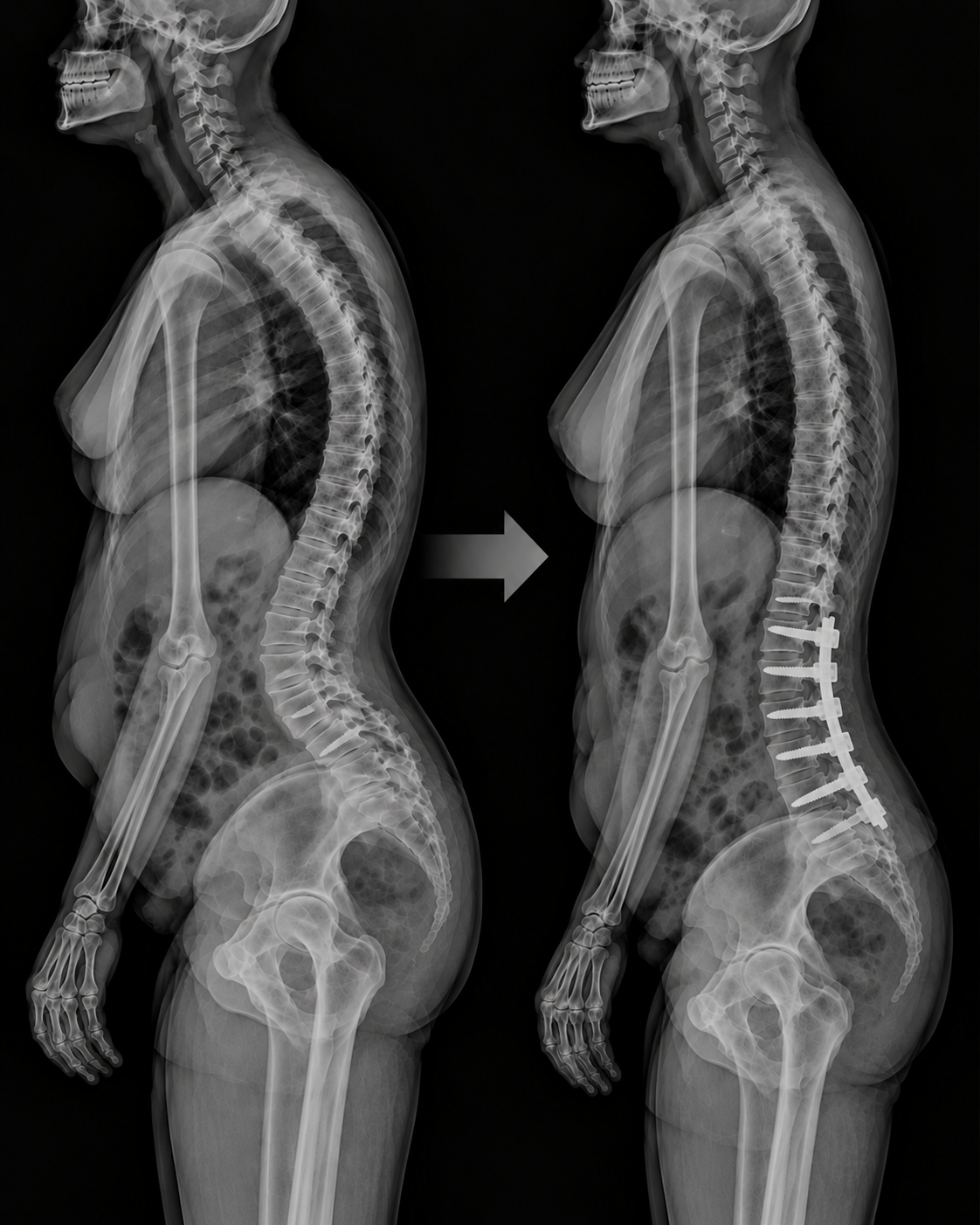
A 356-patient ISSG multicenter study quantifies how thoracic kyphosis changes in response to lumbar correction in adult spinal deformity surgery, identifies three independent predictors of thoracic failure, and makes the case for whole-spine alignment planning.
OPENING SUMMARY
When a surgeon corrects a lumbar spinal deformity, the correction does not stay contained within the fused segments. The thoracic spine above — mobile, unfused, and mechanically connected to everything below — responds. In most patients, that response is beneficial: a flat thoracic spine normalizes, and the whole spinal column moves toward better balance. But in a meaningful proportion of patients, the thoracic response is either insufficient, excessive, or delayed — and over time, it becomes a source of clinical and radiographic failure. This study is the first to systematically quantify both the intended and unintended thoracic changes that follow lumbar correction in adult spinal deformity surgery, tracking them from the early postoperative period through two years of follow-up and identifying the patient and surgical factors that predict when the thoracic spine becomes a problem rather than a partner in recovery.
STUDY SNAPSHOT
Study type – Retrospective multicenter analysis
Number of patients – 356
Surgical procedure – Posterior instrumentation from T9–11 to the pelvis
Minimum follow-up – 2 years
Preoperative TK — normal – 59.8%
Preoperative TK — hypokyphotic – 39.0%
Normalization rate in hypokyphotic patients at 6 weeks – 79.1%
Deterioration rate in normokyphotic patients postoperatively – 11.3%
TK decompensation >15° between 6 weeks and 2 years – 9.3%
Required proximal fusion extension for junctional issues – 9.6%
Total thoracic failure rate – 17.4%
Correlation between TK increase and lumbar lordosis gain – p<0.001; R² = 0.40
PMID – 42760478
DOI – 10.1007/s43390-026-01546-0
Published in – Spine Deformity
Lead author – Renaud Lafage
WHY THIS MATTERS
Adult spinal deformity surgery is fundamentally an alignment restoration procedure — its goal is to bring the spine and pelvis back into balance in a way that reduces pain, improves function, and produces a durable result. Most of the field's planning focus has historically been directed at the lumbar spine, where the deformity is centered: restoring lumbar lordosis, correcting pelvic incidence–lumbar lordosis mismatch, and achieving sagittal vertical axis targets.
But the spine is a kinetic chain. What happens at one level influences what happens at adjacent levels — and the thoracic spine, which typically sits above the upper end of a lumbar deformity fusion, does not simply remain neutral as the lumbar correction unfolds. It participates. The question is whether that participation is therapeutic or pathological.
Prior work has established that reciprocal changes in the thoracic spine occur following lumbar correction. What has been missing is a quantitative characterization of how large those changes are, how reliably they can be predicted from the degree of lumbar correction, how stable they are over time, and which patients are at risk for adverse thoracic outcomes. This study addresses all four questions, providing the most comprehensive published analysis of postoperative thoracic kyphosis morphology in ASD surgery to date.
The clinical stakes are meaningful. Thoracic failure — defined in this study as either significant decompensation or the need for proximal fusion extension — occurred in 17.4% of patients. For a field already managing high rates of mechanical complications and reoperation, understanding and mitigating thoracic failure represents a significant quality improvement opportunity.
UNDERSTANDING THORACIC KYPHOSIS IN ASD
The thoracic spine normally curves gently backward — a kyphotic curve that, in healthy adults, falls within a normative range. In adult spinal deformity patients, this normal curve is often disrupted. Nearly 40% of patients in this cohort were hypokyphotic preoperatively — their thoracic spine was abnormally flat, a pattern often seen in patients who have compensated for lumbar lordosis loss by reducing their thoracic kyphosis in an attempt to maintain overall sagittal balance.
When lumbar lordosis is surgically restored, this compensatory flattening of the thoracic spine is no longer necessary — and the thoracic spine typically responds by increasing its kyphosis back toward normal. This is the reciprocal change that gives the study its central question: how reliable is that response, how large is it, how well does it correlate with what was done in the lumbar spine, and does it last?
The study also distinguishes between two types of thoracic change: iatrogenic change — which occurs because the upper end of the fusion itself directly affects thoracic alignment — and reciprocal change — which occurs in unfused thoracic segments as a mobile response to the correction below. Characterizing both types separately is important for surgical planning, since they reflect different mechanisms and may require different planning responses.
KEY FINDINGS
1. Lumbar correction drove thoracic normalization in most hypokyphotic patients.
Among the 39% of patients with preoperatively hypokyphotic thoracic spines, 79.1% achieved normal thoracic kyphosis by six weeks after surgery. This normalization appeared to be largely a proportional response to lumbar lordosis restoration: multivariate analysis confirmed a significant correlation between postoperative TK increase and lumbar lordosis gain (p<0.001; R² = 0.40), controlling for preoperative alignment and demographics. The R² value of 0.40 indicates that roughly 40% of the variance in postoperative thoracic kyphosis change is explained by the degree of lumbar correction — a meaningful but incomplete relationship, reflecting that other factors also shape the thoracic response.
2. Surgery also created new thoracic abnormalities in some patients.
While surgery normalized most previously flat thoracic spines, it created problems in a subset of patients who had started with normal thoracic curves. Among patients with normal preoperative TK, 11.3% developed abnormal thoracic kyphosis postoperatively. This finding illustrates that the thoracic response is not always beneficial — overcorrection of the lumbar spine, or unfavorable mechanical loading of the proximal junction, can tip an otherwise healthy thoracic curve into pathological territory.
3. Thoracic failure affected nearly 1 in 6 patients by two years.
The composite definition of thoracic failure — either TK decompensation exceeding 15° between 6 weeks and 2 years (9.3% of patients), or requirement for proximal fusion extension due to junctional issues (9.6%) — was met by 17.4% of the cohort. These are not small numbers. Nearly one in six patients undergoing lumbar ASD correction with a fusion ending at T9–T11 experienced a clinically meaningful adverse thoracic outcome within two years.
4. Three independent predictors of thoracic failure were identified.
Multivariate analysis identified three factors independently associated with thoracic failure:
Advanced age (OR 3.6): Older patients were 3.6 times more likely to experience thoracic failure than younger patients. This finding is consistent with evidence across multiple domains of ASD surgery that age represents a fundamental vulnerability — reduced bone quality, greater muscular degeneration, and diminished compensatory reserve all likely contribute.
More proximal lumbar correction (OR 1.9): When the lumbar correction was concentrated in more proximal segments — higher in the lumbar spine — the risk of thoracic failure nearly doubled. This finding has direct surgical planning implications: the location of correction within the lumbar spine, not just the amount, matters for how the thoracic spine responds.
Pronounced early postoperative mid-thoracic kyphosis (OR 2.3): Patients with a pronounced mid-thoracic kyphosis evident at the early postoperative (six-week) assessment were 2.3 times more likely to experience thoracic failure by two years. This early imaging signal is particularly important because it suggests that the six-week postoperative radiograph is not just a confirmatory check — it may be a prognostically meaningful window for identifying patients at elevated risk before failure becomes established.
5. The relationship between thoracic and lumbar changes was bidirectional and dynamic.
The finding that TK increase correlated with lumbar lordosis gain, while informative, captures only part of the story. Between six weeks and two years — a period when fusion consolidation is occurring and the mechanical environment is changing — 9.3% of patients experienced TK decompensation exceeding 15°. The thoracic spine's response to lumbar correction is not a one-time event that stabilizes at the first postoperative visit; it continues to evolve over the first two years and requires longitudinal monitoring to characterize fully.
THE THORACIC FAILURE FRAMEWORK: TWO DISTINCT PATHWAYS
The study's composite definition of thoracic failure captures two distinct clinical presentations that merit separate consideration:
TK Decompensation (9.3% of patients)
This pathway involves progressive increase in thoracic kyphosis beyond the six-week postoperative value, exceeding a threshold of 15° of change. Decompensation of this magnitude represents a meaningful loss of thoracic alignment — one that can shift the overall sagittal balance, increase proximal junctional stress, and potentially lead to clinical symptoms. It likely reflects progressive collapse or settling at the proximal junction of the lumbar fusion construct, or loss of muscular support for thoracic posture in older patients with limited physiological reserve.
Proximal Fusion Extension for Junctional Issues (9.6% of patients)
This pathway involves a return to the operating room to extend the fusion construct further up the thoracic spine to address junctional kyphosis that has become clinically symptomatic or radiographically severe enough to warrant surgical intervention. This is the more consequential outcome — it represents a second major surgical procedure with its own risks, recovery demands, and resource utilization.
The overlap between these two pathways is not specified in the abstract, but together they define a 17.4% thoracic failure rate that is clinically meaningful and substantially underappreciated in ASD surgical planning.
PRACTICAL IMPLICATIONS
Preoperative thoracic kyphosis morphology should inform surgical planning — not just be documented.
The finding that the location and magnitude of lumbar correction predicts the thoracic response means that thoracic kyphosis assessment before surgery is not merely descriptive — it is actionable. Surgeons planning lumbar ASD correction should evaluate preoperative TK in the context of planned lumbar lordosis restoration and consider how that correction is distributed across lumbar segments. For patients who start with pronounced mid-thoracic kyphosis or who are older, the planning conversation should include specific attention to proximal junction management.
The six-week postoperative radiograph is prognostically important for thoracic outcomes.
The identification of pronounced early postoperative mid-thoracic kyphosis as an independent predictor of two-year failure means that the six-week visit is not just a routine early check — it is a risk stratification opportunity. Patients whose six-week thoracic shape is concerning should be identified early and monitored with increased frequency, as their risk of progressive decompensation or proximal junctional failure is substantially elevated.
More proximal lumbar correction carries higher thoracic failure risk.
The association between proximal lumbar correction location and thoracic failure has direct implications for how surgeons distribute their correction across lumbar segments. When correction is concentrated more proximally in the lumbar spine — closer to the thoracolumbar junction — the mechanical loading on the thoracic spine and proximal junction is altered in ways that appear to increase failure risk. Distributing correction more distally, when anatomically feasible, may reduce this risk.
Age should explicitly inform thoracic failure risk counseling.
At an odds ratio of 3.6 for advanced age, the age-thoracic failure association is among the strongest predictors in the study. Older patients undergoing lumbar ASD correction with a fusion ending at T9–T11 should be counseled about the approximately threefold higher risk of adverse thoracic outcomes, and their postoperative surveillance plan should be designed accordingly.
Tailored alignment strategies may reduce thoracic failure rates.
The study's conclusion explicitly calls for tailored alignment strategies for patients with the identified risk factors. The concept of one-size-fits-all lumbar correction targets — applied uniformly regardless of patient age, preoperative thoracic shape, and planned correction location — appears insufficient when 17.4% of patients experience thoracic failure within two years. For high-risk patients, this might mean planning more aggressive proximal fusion to begin with, selecting alignment targets that minimize the thoracic response, or implementing enhanced postoperative surveillance that enables earlier intervention when decompensation is detected.
The R² of 0.40 signals room for improvement in predicting thoracic response.
While the correlation between lumbar lordosis gain and TK increase is statistically significant and clinically meaningful, an R² of 0.40 means that 60% of the variance in thoracic response is not explained by lumbar correction magnitude alone. Future research should investigate what additional factors — including thoracic muscular strength and quality, preoperative thoracic flexibility, bone quality, and the specific distribution of lumbar correction — account for the remainder. Better predictive models would enable more individualized surgical planning.
CONCLUSION
This study establishes a clear and clinically actionable principle: in adult spinal deformity surgery with fusion ending between T9 and T11, the thoracic spine responds to lumbar correction in a largely proportional and often beneficial way — but nearly one in six patients experiences thoracic failure within two years, and that failure is predictable from preoperative and early postoperative factors that are available at the time of surgical planning and early follow-up. Advanced age, more proximal lumbar correction, and pronounced early mid-thoracic kyphosis independently identify patients at elevated risk. For the field, this means that whole-spine alignment planning — explicitly accounting for the anticipated thoracic response and the risk factors for adverse thoracic outcomes — is not optional. The thoracic spine is not a passive bystander in lumbar deformity surgery. It is an active participant whose response determines, in part, whether the operation succeeds in the long run.
READ THE FULL STUDY