Your Spine X-Ray May Reveal More Than Your Spine: Muscle Loss Detection in Adult Spinal Deformity
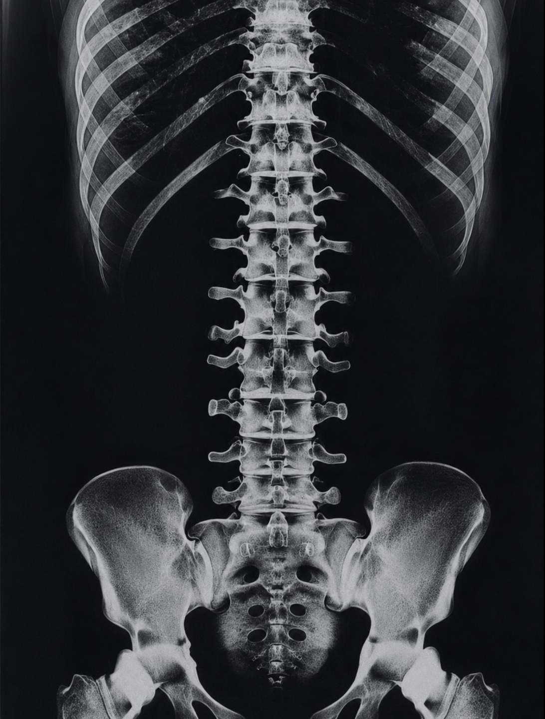
A 24-center North American study finds that routine full-body radiographs used in spinal deformity care can identify sarcopenia — at no additional cost or radiation — and that muscle loss reshapes how the entire body compensates for spinal imbalance.
OPENING SUMMARY
Sarcopenia — the progressive loss of muscle mass and strength associated with aging — is a known risk factor for worse surgical outcomes in patients with adult spinal deformity. But it is rarely measured systematically before surgery, in part because doing so typically requires dedicated testing beyond what is already part of standard care. This study challenges that assumption. By measuring thigh and quadriceps muscle thickness directly from full-body radiographs already obtained as part of routine ASD workup, researchers demonstrated that sarcopenia can be identified opportunistically — without additional imaging, cost, or radiation exposure to the patient. And what that measurement reveals about a patient's overall health and postural compensation strategy turns out to be clinically significant.
STUDY SNAPSHOT
Study type – Multicenter retrospective cohort study of prospectively collected data
Number of patients – 540
Number of centers – 24 U.S. and Canadian spine centers
Data collection period– 2019–2024
Mean age – 60 years
Female – 71%
Sarcopenic patients – 61 (11.3%)
Sarcopenia definition – Both AP thigh and LAT quadriceps measurements below validated sex-specific full-body cutoffs
Clinical comparators – Frailty scores, grip strength, 3-meter timed up and go (TUG), epigenetic age
Radiographic comparators – Thoracic kyphosis, cervical lordosis, sagittal vertical axis, knee flexion angle, pelvic tilt, pelvic shift, sacro-femoral angle
Level of evidence – Prognostic Level III
WHY THIS MATTERS
Adult spinal deformity patients are disproportionately older, frequently frail, and often arrive at surgical evaluation carrying a burden of comorbidities that standard imaging does not capture. Sarcopenia is one of the most clinically important of these — it is associated with impaired recovery, increased complication risk, and worse postoperative functional outcomes. Yet identifying it typically requires tests that fall outside the routine spine surgery workup.
full-body radiography is already standard of care for ASD patients, providing standing images from head to foot that allow precise measurement of spinal and pelvic alignment. The insight behind this study is straightforward but consequential: if those images already include the thighs, why not measure the muscles too?
If thigh muscle measurements from routine full-body imaging can reliably identify sarcopenic patients — and if that identification predicts clinically meaningful differences in frailty, functional performance, and postural compensation — then a powerful new screening tool may already be sitting in the hands of every spine surgeon who orders a standard full-body radiograph.
KEY FINDINGS
1. Sarcopenia was present in roughly 1 in 9 ASD patients. Using validated sex-specific thresholds applied to full-body-derived thigh and quadriceps measurements, 11.3% of the 540-patient cohort met criteria for sarcopenia. Sarcopenic patients had meaningfully lower BMI (23.6 vs. 27.3 kg/m²), higher clinical frailty scores (3.4 vs. 3.0), and slower timed up-and-go performance (12.2 vs. 10.5 seconds) compared to non-sarcopenic patients — all statistically significant differences.
2. Full-body-derived muscle measurements correlated with multiple validated markers of physical decline. On multivariate analysis, smaller thigh and quadriceps thickness — as measured from full-body imaging — independently correlated with higher frailty scores, weaker grip strength, slower TUG performance, and older epigenetic age. Epigenetic age — a biological marker derived from cellular aging patterns that can differ from chronological age — adds a particularly novel dimension to these findings, suggesting that muscle loss as detected on imaging reflects not just physical function but the pace of biological aging itself.
3. Sarcopenia changed how the body compensated for spinal imbalance. Sarcopenic patients showed a distinct compensatory postural profile compared to non-sarcopenic patients. They demonstrated greater thoracic kyphosis, greater cervical lordosis, a larger sagittal vertical axis, and greater knee flexion angle — all statistically significant associations. Notably, proximal compensatory mechanisms involving the pelvis — pelvic tilt, pelvic shift, and sacro-femoral angle — were not significantly associated with sarcopenia. This pattern suggests that sarcopenic patients rely more heavily on distal compensations (knees, cervical spine) while proximal pelvic mechanisms are relatively impaired — a finding with potential implications for surgical planning and postoperative recovery expectations.
4. The measurement requires no additional imaging, cost, or radiation. Because thigh and quadriceps thickness can be derived from full-body radiographs already obtained as standard of care, this screening approach adds no burden to the patient and no additional cost to the workup. The authors describe this as an opportunistic measurement — one that extracts additional clinically useful information from imaging that would be performed regardless.
PRACTICAL IMPLICATIONS
Sarcopenia screening may be within reach for every ASD patient who undergoes full-body imaging. The opportunistic nature of this measurement is its most immediately practical feature. If validated further, incorporating thigh and quadriceps measurements into routine full-body analysis could become a low-barrier way to flag sarcopenic patients before surgery — without adding a separate referral, test, or appointment.
Identifying sarcopenia preoperatively opens the door to targeted intervention. Knowing that a patient is sarcopenic before surgery creates an opportunity to address it — through prehabilitation, nutritional support, or physical therapy aimed at building muscle mass and functional reserve before an elective procedure. Whether such interventions improve ASD surgical outcomes is a natural next question for future research, but the first step is reliable identification.
Sarcopenic patients may need different surgical planning assumptions. The finding that sarcopenic patients compensate differently for sagittal imbalance — relying more on distal mechanisms like knee flexion rather than pelvic response — has potential implications for how surgeons anticipate postoperative alignment behavior and recovery in this subgroup. A patient whose body compensates primarily through the knees may respond differently to spinal realignment than one whose pelvis is the primary compensatory mechanism.
Epigenetic age as an outcome variable adds a novel dimension. The association between full-body-derived muscle measurements and epigenetic age suggests that this imaging-based tool may be capturing something deeper than muscle size alone — it may reflect the biological aging process more broadly. This is an early-stage finding that warrants further investigation but adds scientific interest to the clinical utility of the approach.
CONCLUSION
This study makes a practical and scientifically grounded case for incorporating thigh muscle measurement into the routine interpretation of full-body radiographs in adult spinal deformity patients. By demonstrating that these measurements correlate with validated markers of frailty, functional performance, and biological aging — and that sarcopenic patients compensate for spinal imbalance in a distinct and clinically relevant way — the authors have identified a low-cost, zero-burden screening opportunity that could meaningfully improve preoperative risk stratification and surgical planning. The next step for the field is to determine whether acting on that information, through prehabilitation or other targeted interventions, translates into better surgical outcomes for this vulnerable patient population.
READ THE FULL STUDY