Preventing the Spine From Collapsing Above the Fusion: A New Framework for Upper Instrumented Vertebra Selection in Adult Spinal Deformity Surgery
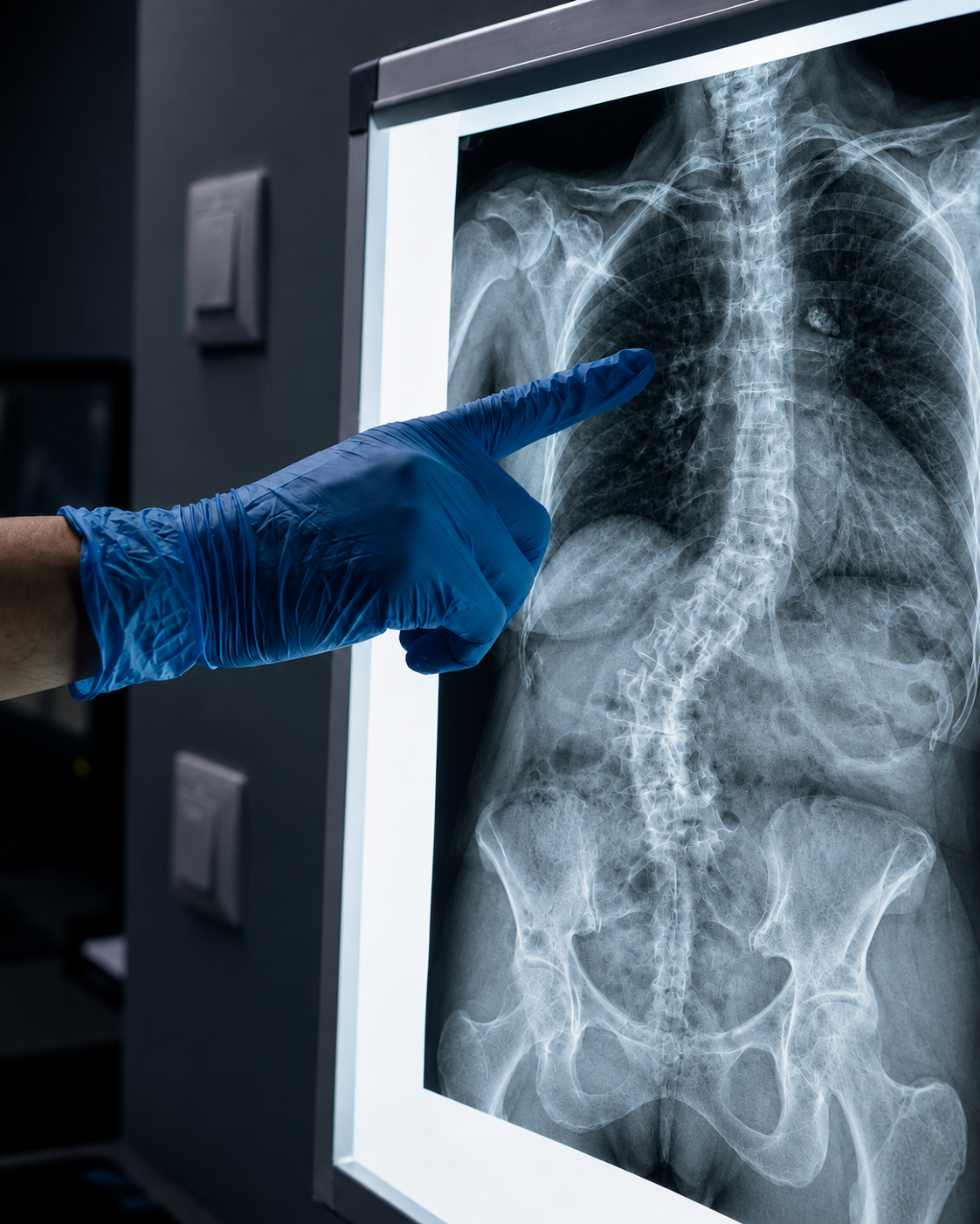
A 627-patient, 20-center study finds that preoperative cervicothoracic alignment predicts proximal junctional kyphosis risk — and that extending the fusion to the upper thoracic spine reduces that risk by 36% in patients with worse baseline alignment.
OPENING SUMMARY
Proximal junctional kyphosis — a progressive collapse of the spine just above where a fusion construct ends — is one of the most consequential complications in adult spinal deformity surgery. It can erode the benefits of an otherwise successful operation, cause significant pain and neurological symptoms, and in its most severe form require major revision surgery. Despite its clinical importance, objective guidance for one of the key decisions that influences PJK risk — where to end the fusion — has been largely absent from the literature. Surgeons have relied primarily on experience and judgment to select the upper instrumented vertebra, the topmost level included in a spinal fusion construct. This study changes that, providing the first data-driven framework for UIV selection based on preoperative alignment measurement — and quantifying how much that decision matters for patient outcomes.
STUDY SNAPSHOT
Study type – Multicenter prospective ASD registry analysis
Number of patients – 627
Number of centers – 20
Median age – 66 years (IQR 59–70)
Female – 483 (77%)
Lower thoracic UIV (T9–L1) – 380 patients (61%)
Upper thoracic UIV (T1–T5) – 247 patients (39%)
Minimum follow-up – 2 years radiological follow-up
Primary outcome – Proximal junctional kyphosis within 2 years
Key preoperative measure – C2–T9 pelvic angle (PA)
Cohort median C2–T9 PA – 14°
Published in – The Bone & Joint Journal, Volume 108-B, Issue 7 (July 2026), pages 943–951
PMID – 42379558
DOI – 10.1302/0301-620X.108B7.BJJ-2025-1405.R1
WHY THIS MATTERS
Adult spinal deformity surgery is among the most technically demanding procedures in orthopedic and neurological surgery. Fusion constructs typically extend from the thoracic spine down to the sacrum or pelvis — long, complex constructs that require precise planning at every level. One of the most consequential planning decisions is selecting the upper instrumented vertebra: the topmost vertebra included in the fusion. Stop too low, and the construct may fail at its upper end through proximal junctional kyphosis. Stop too high, and the surgery becomes more extensive, with greater potential for complications, longer recovery, and more levels of spinal motion sacrificed.
Proximal junctional kyphosis occurs when the spine kyphoses — bends forward — at the junction just above the fusion, where the rigid instrumented construct meets the mobile native spine. This transition zone is subject to significant mechanical stress, and when it fails, it can do so progressively and painfully. PJK is not a minor inconvenience: in its most severe manifestation — proximal junctional failure — it can cause neurological compromise and necessitate major revision surgery.
Despite extensive research into the factors that influence PJK, objective methods for UIV selection have been lacking. This study addresses that gap directly, combining preoperative alignment measurement with a large multicenter dataset to establish the first evidence-based framework for where to end the fusion — and for whom extending to the upper thoracic spine provides the greatest benefit.
UNDERSTANDING THE KEY MEASUREMENT: C2–T9 PELVIC ANGLE
The C2–T9 pelvic angle is a radiographic measurement that captures the relationship between the cervicothoracic spine and the pelvis — reflecting the degree of sagittal malalignment in the upper portion of the spinal column. Unlike measurements that focus on lumbar or global alignment alone, the C2–T9 PA specifically captures alignment in the region just above where a lower thoracic fusion would end — making it particularly relevant to the mechanical forces acting on the proximal junction of a fusion construct.
A higher C2–T9 PA reflects worse cervicothoracic malalignment. Because this measurement can be derived from standard preoperative imaging, it is practically accessible at the time of surgical planning — making it an actionable tool rather than a research construct.
KEY FINDINGS
1. PJK was dramatically more common with lower thoracic fusion endpoints. Among the 380 patients whose fusion ended in the lower thoracic spine (T9–L1), PJK developed in 149 — a rate of 39% within two years. Among the 247 patients with an upper thoracic UIV (T1–T5), only 38 developed PJK — a rate of 15%. This nearly threefold difference in PJK rates between UIV regions is one of the most striking findings in the study and establishes the stakes of the UIV selection decision clearly.
2. The relationship between preoperative alignment and PJK risk depended significantly on UIV region. A significant interaction was identified between preoperative C2–T9 PA and UIV region (p = 0.028), meaning that the effect of preoperative alignment on PJK risk was not the same for lower and upper thoracic fusion endpoints. Worse preoperative cervicothoracic alignment was associated with higher PJK risk — but this relationship was modified by where the fusion ended. Patients with worse alignment who received a lower thoracic UIV were at particularly elevated risk, while those who received an upper thoracic UIV appeared to be partially protected.
3. Extending the fusion to the upper thoracic spine reduced PJK risk by 36% at the cohort's median alignment. At a preoperative C2–T9 PA of 14° — the median value for the cohort — an upper thoracic UIV was associated with an adjusted absolute risk reduction of 36% compared to a lower thoracic UIV. The number needed to expose (NNEB) was 2.8, meaning that for every 2.8 patients who receive an upper thoracic UIV instead of a lower thoracic UIV, one case of PJK is prevented. This is a clinically meaningful effect size that justifies serious consideration of UIV extension in appropriately identified patients.
4. Female sex was an independent predictor of PJK. After adjusting for alignment and UIV region, female sex was independently associated with a 62% higher odds of developing PJK (OR 1.62; 95% CI 1.03–2.59; p = 0.042). In a surgical population that is 77% female, this finding has broad applicability and supports the inclusion of sex as a variable in PJK risk stratification. The biological mechanisms underlying this association — which may include differences in bone density, spinal morphology, and compensatory strategies — warrant further investigation.
5. The framework focuses on factors available at the time of surgical planning. A deliberate strength of this study's design is its focus on preoperative factors — specifically the C2–T9 PA, which is measurable from standard imaging obtained before surgery. This makes the framework immediately applicable in clinical practice: surgeons can measure the C2–T9 PA during preoperative planning, assess deformity severity and sex, and use that information to inform UIV selection before the patient arrives in the operating room.
PRACTICAL IMPLICATIONS
UIV selection should be integrated into preoperative alignment analysis, not determined during surgery. The most important practical implication of this study is that where a fusion ends should be a deliberate, data-informed preoperative decision — not an intraoperative judgment call made under time pressure. Incorporating C2–T9 PA measurement into the standard preoperative radiographic workup gives surgeons an objective basis for UIV selection that complements clinical experience and individual patient factors.
Patients with worse preoperative cervicothoracic alignment are the strongest candidates for upper thoracic UIV extension. The interaction between C2–T9 PA and UIV region means that the benefit of extending the fusion is not uniform across all patients — it is greatest in those with worse preoperative alignment. This is clinically useful: rather than recommending upper thoracic UIV for all patients, the framework identifies a specific alignment-based criterion for selecting those most likely to benefit from a more extensive fusion.
Sex-specific risk counseling should be standard in ASD surgical planning. The independent association between female sex and PJK risk — in a population that is three-quarters female — means that sex should be explicitly incorporated into preoperative risk conversations. Female patients, particularly those with elevated C2–T9 PA, may warrant particular attention to UIV selection and postoperative monitoring for junctional failure.
The number needed to expose of 2.8 provides a practical benchmark for clinical decision-making. An NNEB of 2.8 means that extending the fusion to the upper thoracic spine in fewer than three patients at median alignment levels would prevent one case of PJK. When weighed against the consequences of PJK — including pain, loss of correction, and potentially major revision surgery — this is a favorable tradeoff for many patients. Individual decisions will still depend on patient-specific factors including age, comorbidities, bone quality, and surgical risk tolerance.
This framework is a starting point, not a definitive algorithm. UIV selection in ASD surgery is multifactorial, and no single measurement should be used in isolation. The C2–T9 PA is an important and accessible tool, but it should be integrated with other alignment parameters, patient-specific risk factors, bone quality assessment, and surgeon expertise. The study's authors frame this as a framework for UIV selection — a structured approach to a complex decision — rather than a binary rule.
CONCLUSION
This study makes a meaningful and practically useful contribution to one of the most persistent challenges in adult spinal deformity surgery: how to decide where to end the fusion. By demonstrating that preoperative C2–T9 pelvic angle predicts PJK risk, that this risk interacts significantly with UIV region, and that extending the fusion to the upper thoracic spine reduces absolute PJK risk by 36% at median alignment values, the authors have provided the field with an evidence-based foundation for a decision that has long depended primarily on intuition and experience. Combined with the independent finding that female sex increases PJK risk by 62%, this study supports a more individualized, data-informed approach to UIV selection — one that could meaningfully reduce one of ASD surgery's most consequential complications.
READ THE FULL STUDY