How the Body Compensates for a Failing Spine: Four Distinct Patterns — and Why They Matter for Surgical Planning
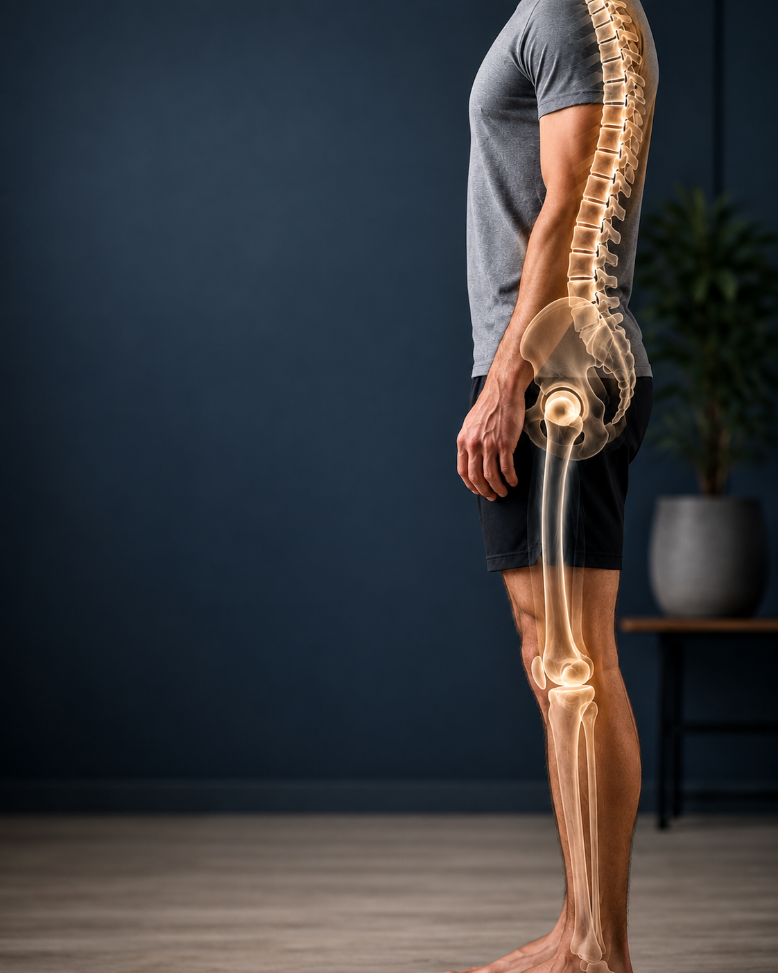
A multicenter study of 871 adult spinal deformity patients identifies four clinically distinct patterns of lower limb compensation using cluster analysis, revealing that how a patient compensates for spinal imbalance is shaped by anatomy, sex, joint disease, frailty, and deformity severity.
OPENING SUMMARY
When the spine loses its ability to maintain upright sagittal balance, the body does not simply collapse — it adapts. The hips extend, the knees flex, the pelvis shifts, and the ankles adjust in coordinated compensatory strategies that allow patients to remain standing despite severe spinal malalignment. This compensation has long been recognized as a feature of adult spinal deformity — but it has been treated largely as a single phenomenon. In reality, not all patients compensate the same way, and the differences between them may carry important clinical information. This study is among the first to systematically characterize the different patterns of lower limb compensation in ASD patients, identifying four distinct compensatory clusters and describing how each is shaped by a unique combination of patient and disease characteristics. The findings open a new dimension of preoperative assessment — one that looks beyond spinal alignment to the whole-body strategies patients have developed to live with it.
STUDY SNAPSHOT
Study type – Multicenter prospective database analysis
Number of patients – 871
Female – 66.9%
Mean age – 62.3 ± 14.6 years
Mean BMI – 27.7 ± 5.5 kg/m²
Imaging – Full-body X-rays in erect position at baseline
Key radiographic measures – Sacro-femoral angle (SFA), knee flexion angle (KA), pelvic incidence (PI), PI-LL mismatch, T1 pelvic angle (TPA), pelvic shift, ankle dorsiflexion angle
Cluster analysis method – K-means clustering; optimal cluster number determined by silhouette score
Number of clusters identified – 4
Clinical measures – Disability scores, frailty scores, SRS scores, hip and knee OA grade
Published in – European Spine Journal, Volume 35, Issue 5, May 2026, pages 2324–2334
PMID – 41555093
DOI – 10.1007/s00586-026-09742-2
WHY THIS MATTERS
Sagittal malalignment in adult spinal deformity creates a forward imbalance — the body's center of mass shifts in front of its support base. To counteract this, the lower limbs recruit compensatory mechanisms: the pelvis tilts backward (retroversion), the hips extend, the knees flex, and the ankles dorsiflex. These adaptations allow patients to maintain an upright posture despite a deformed spine, but they do so at a physiological cost — placing abnormal mechanical demands on the joints and muscles of the lower extremities, contributing to pain, fatigue, and functional limitation.
The clinical importance of lower limb compensation has been recognized in ASD research for some time. Studies have shown that compensatory mechanisms — particularly knee flexion — are associated with worse patient-reported outcomes and higher disability scores. But the field has largely treated compensation as a single variable: patients either compensate or they do not, and the degree of compensation is measured by individual parameters like pelvic tilt or knee flexion angle.
What has been missing is a systematic characterization of whether different patients recruit fundamentally different compensatory strategies — and if so, whether those strategies reflect distinct clinical profiles with different implications for surgical planning, outcome prediction, and postoperative course. This study addresses that gap directly, using unsupervised cluster analysis to let the data reveal natural groupings rather than imposing predetermined categories.
UNDERSTANDING THE KEY MEASUREMENTS
Sacro-femoral angle (SFA) measures the relationship between the sacrum and the femur, reflecting the degree of hip extension used in compensation. A more negative SFA indicates greater hip extension.
Knee flexion angle (KA) measures the degree of knee bending used to compensate for forward imbalance. Greater knee flexion is associated with higher energy expenditure and greater joint loading.
Pelvic incidence (PI) is a fixed anatomical parameter that describes the relationship between the sacrum and the pelvis. It does not change with posture and serves as a morphological baseline that influences how much lumbar lordosis a given patient needs and what compensatory strategies are most accessible to them.
PI-LL mismatch is the difference between pelvic incidence and lumbar lordosis — a key measure of sagittal deformity severity. A large mismatch indicates significant spinal imbalance.
T1 pelvic angle (TPA) is a global measure of sagittal alignment that captures the overall forward shift of the upper body relative to the pelvis. Higher values indicate more severe global malalignment.
THE FOUR COMPENSATORY PATTERNS
No Compensators Patients in this cluster showed minimal recruitment of lower limb compensatory mechanisms. They had the lowest PI-LL mismatch (5.3° ± 21.3°) and the lowest TPA values (16.1° ± 11.5°), indicating relatively preserved spinal alignment with limited demand on lower limb compensation. This group represents patients whose spinal deformity has not yet — or has not sufficiently — overwhelmed the spine's intrinsic capacity for balance.
Recliners (Primarily Hip Extension) This cluster was characterized by compensation primarily through hip extension — extending the hips backward to shift the pelvis and counteract forward trunk lean. Recliners had the highest proportion of female patients (79.3%) of any cluster, the highest pelvic incidence values (mean 59.2°), and the lowest rate of severe hip osteoarthritis (38.5%). The high pelvic incidence in this group may facilitate hip extension as a compensatory strategy, as greater pelvic incidence is associated with greater potential for pelvic retroversion. The relative preservation of hip joint health in this group may also enable this strategy to be recruited effectively.
Squatters (Primarily Knee Flexion) This cluster was characterized by compensation primarily through knee flexion — a strategy associated with significantly higher physiological burden. Squatters had the highest rates of both hip OA (71.9%) and knee OA (72.7%), the worst disability scores, the worst frailty scores, and the poorest SRS quality-of-life scores of any group. They had the lowest proportion of female patients (47.0%) and significantly higher BMI than other clusters. The reliance on knee flexion in this group may reflect limited capacity to recruit hip extension — potentially due to severe hip OA — and the physiological costs of sustained knee flexion may contribute to the worse functional profile observed.
Mixed Compensators (Both Hip Extension and Knee Flexion) Patients in this cluster recruited both hip extension and knee flexion simultaneously — suggesting that neither strategy alone was sufficient to compensate for their degree of malalignment. Mixed compensators had the greatest PI-LL mismatch (30.4° ± 20.0°) and the highest TPA values (33.3° ± 11.7°), indicating the most severe sagittal imbalance of any cluster. They also had significantly higher BMI. The recruitment of multiple compensatory strategies in this group likely reflects a greater overall demand placed on the lower extremities by more advanced spinal deformity.
KEY FINDINGS
1. Four distinct compensatory patterns exist in ASD patients — not a single phenomenon. K-means cluster analysis identified four stable, clinically distinct compensatory patterns, confirmed by silhouette scoring as the optimal solution. This finding establishes that lower limb compensation in ASD is heterogeneous and that treating it as a single variable misses important clinical differentiation.
2. Squatters bore the greatest overall clinical burden. The knee flexion-dominant group was distinguished by the worst functional and quality-of-life scores, the highest rates of both hip and knee OA, the highest frailty burden, and significantly higher BMI. This cluster represents the most physiologically taxed patients in the cohort — those for whom compensation is both most necessary and most costly.
3. Sex, pelvic anatomy, and joint disease were major determinants of compensatory strategy. The strong female predominance in Recliners (79.3%) versus the male-skewed distribution in Squatters (47.0% female) suggests that biological sex influences which compensatory strategy is recruited — potentially through differences in pelvic morphology, muscle mass, or joint disease patterns. The strong association between hip OA and knee flexion-dominant compensation suggests that joint disease in the hips limits the capacity to recruit hip extension, forcing recruitment of knee flexion instead — a strategy that then further loads the already-compromised knee joint.
4. Deformity severity drove the recruitment of multiple compensatory mechanisms. Mixed compensators — those using both hip extension and knee flexion — had the most severe sagittal imbalance by both PI-LL mismatch and TPA. This gradient across clusters (No Compensators → Recliners → Squatters/Mixed → greater severity) suggests that compensation escalates as deformity worsens, with more mechanisms recruited as individual strategies prove insufficient.
5. Pelvic incidence shaped the compensatory landscape. Recliners and Mixed compensators had the highest pelvic incidence values, while No Compensators and Squatters had lower values. This finding is consistent with prior evidence that higher pelvic incidence facilitates greater pelvic retroversion — and suggests that pelvic anatomy is an important determinant of which compensatory strategies are available to a given patient.
PRACTICAL IMPLICATIONS
Preoperative assessment should include characterization of lower limb compensatory patterns. Understanding which compensatory strategy a patient is using before surgery has potential implications for surgical planning, outcome prediction, and postoperative management. A patient who is a Squatter — with high knee and hip OA burden, high frailty, and poor functional scores — represents a fundamentally different surgical risk profile and recovery trajectory than a Recliner, even if both have similar global alignment parameters on X-ray.
Squatters may require particular attention to comorbidity management and functional rehabilitation. The clustering of adverse characteristics in the Squatter group — high OA burden, high frailty, worst functional scores — suggests that this subgroup may benefit from more intensive preoperative optimization, including attention to joint health, frailty mitigation, and functional conditioning before surgery. Their postoperative course may also differ from other groups in ways that warrant closer monitoring.
Surgical correction may not fully address lower limb compensation if joint disease limits recovery. If a patient's compensatory knee flexion is driven partly by severe hip or knee OA rather than purely by spinal imbalance, correcting the spinal deformity may not fully resolve the compensatory posture. In such patients, orthopedic evaluation of the hip and knee — and potential joint replacement — may be part of comprehensive treatment planning. This study supports a whole-body, multi-specialty approach to ASD care.
The four-cluster framework may eventually inform individualized surgical planning and outcome prediction. If future studies validate these compensatory patterns as stable, reproducible categories with distinct outcome trajectories, they could become a useful classification tool in clinical practice — helping surgeons anticipate which patients will and will not fully resolve their compensatory posture after spinal correction, and counseling patients accordingly.
The finding that compensation is multifactorial reinforces the need for whole-body radiographic assessment. Full-body standing X-rays — which capture not just the spine but the hips, knees, and ankles — are essential for characterizing compensatory patterns. This study's findings support the clinical value of full-body imaging in ASD assessment, as standard lumbar or thoracolumbar films would not capture the lower limb parameters needed to identify compensatory type.
CONCLUSION
This study makes a compelling case that lower limb compensation in adult spinal deformity is not a single, uniform phenomenon — it is a heterogeneous set of strategies shaped by pelvic anatomy, joint disease, sex, body composition, frailty, and deformity severity. The four compensatory patterns identified — No Compensators, Recliners, Squatters, and Mixed Compensators — each carry distinct clinical profiles with different implications for how we understand, plan for, and manage ASD patients. The Squatter profile in particular — with its convergence of high OA burden, frailty, disability, and poor functional scores — deserves focused clinical attention as a high-risk subgroup. More broadly, this research supports a paradigm shift in ASD assessment: from spine-centric analysis toward a whole-body understanding of how patients are living with and compensating for their deformity before surgery ever begins.
READ THE FULL STUDY